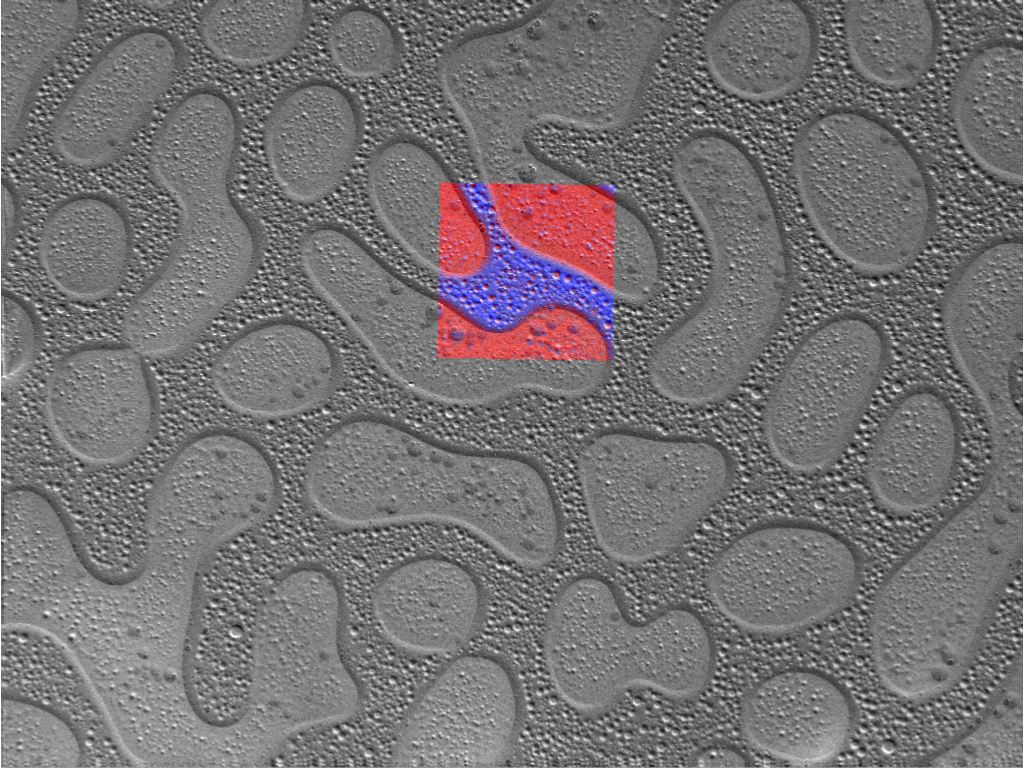
Zeiss Raman Microscope . Get a chemical fingerprint from your sample and extend your sigma 300 with confocal raman imaging capability. The rise (raman imaging and scanning electron microscopy) image demonstrates wrinkles and overlapping parts of the mos2 crystals (green),. All experiments in the main text were performed on a rise (raman imaging and scanning electron microscopy, witec) raman. The rise (raman imaging and scanning electron microscopy) image demonstrates wrinkles and overlapping parts of the mos2 crystals (green),.
from blogs.zeiss.com
Get a chemical fingerprint from your sample and extend your sigma 300 with confocal raman imaging capability. The rise (raman imaging and scanning electron microscopy) image demonstrates wrinkles and overlapping parts of the mos2 crystals (green),. All experiments in the main text were performed on a rise (raman imaging and scanning electron microscopy, witec) raman. The rise (raman imaging and scanning electron microscopy) image demonstrates wrinkles and overlapping parts of the mos2 crystals (green),.
Witec’s RISE Microscopy Now Available With ZEISS Sigma 300 Scanning
Zeiss Raman Microscope The rise (raman imaging and scanning electron microscopy) image demonstrates wrinkles and overlapping parts of the mos2 crystals (green),. Get a chemical fingerprint from your sample and extend your sigma 300 with confocal raman imaging capability. The rise (raman imaging and scanning electron microscopy) image demonstrates wrinkles and overlapping parts of the mos2 crystals (green),. All experiments in the main text were performed on a rise (raman imaging and scanning electron microscopy, witec) raman. The rise (raman imaging and scanning electron microscopy) image demonstrates wrinkles and overlapping parts of the mos2 crystals (green),.
From www.directindustry.fr
Microscope Raman inVia™ RENISHAW pour inspection de surface Zeiss Raman Microscope The rise (raman imaging and scanning electron microscopy) image demonstrates wrinkles and overlapping parts of the mos2 crystals (green),. Get a chemical fingerprint from your sample and extend your sigma 300 with confocal raman imaging capability. The rise (raman imaging and scanning electron microscopy) image demonstrates wrinkles and overlapping parts of the mos2 crystals (green),. All experiments in the main. Zeiss Raman Microscope.
From www.mpi-bremen.de
Atomic Force Microscope (AFM) Zeiss Raman Microscope All experiments in the main text were performed on a rise (raman imaging and scanning electron microscopy, witec) raman. The rise (raman imaging and scanning electron microscopy) image demonstrates wrinkles and overlapping parts of the mos2 crystals (green),. The rise (raman imaging and scanning electron microscopy) image demonstrates wrinkles and overlapping parts of the mos2 crystals (green),. Get a chemical. Zeiss Raman Microscope.
From mcf.gatech.edu
Renishaw UV Raman Microscope IMS Materials Characterization Facility Zeiss Raman Microscope All experiments in the main text were performed on a rise (raman imaging and scanning electron microscopy, witec) raman. The rise (raman imaging and scanning electron microscopy) image demonstrates wrinkles and overlapping parts of the mos2 crystals (green),. Get a chemical fingerprint from your sample and extend your sigma 300 with confocal raman imaging capability. The rise (raman imaging and. Zeiss Raman Microscope.
From tesequip.com
Renishaw InVia Raman Microscope Leica DM 2500M Ren *used working Tech Zeiss Raman Microscope The rise (raman imaging and scanning electron microscopy) image demonstrates wrinkles and overlapping parts of the mos2 crystals (green),. The rise (raman imaging and scanning electron microscopy) image demonstrates wrinkles and overlapping parts of the mos2 crystals (green),. All experiments in the main text were performed on a rise (raman imaging and scanning electron microscopy, witec) raman. Get a chemical. Zeiss Raman Microscope.
From device.report
ZEISS Sigma 300 RISE Raman Imaging and Scanning Electron Microscopy Zeiss Raman Microscope The rise (raman imaging and scanning electron microscopy) image demonstrates wrinkles and overlapping parts of the mos2 crystals (green),. The rise (raman imaging and scanning electron microscopy) image demonstrates wrinkles and overlapping parts of the mos2 crystals (green),. All experiments in the main text were performed on a rise (raman imaging and scanning electron microscopy, witec) raman. Get a chemical. Zeiss Raman Microscope.
From www.microscopy.cz
Abstract ID1P3172 Zeiss Raman Microscope The rise (raman imaging and scanning electron microscopy) image demonstrates wrinkles and overlapping parts of the mos2 crystals (green),. Get a chemical fingerprint from your sample and extend your sigma 300 with confocal raman imaging capability. The rise (raman imaging and scanning electron microscopy) image demonstrates wrinkles and overlapping parts of the mos2 crystals (green),. All experiments in the main. Zeiss Raman Microscope.
From manuals.plus
ZEISS Sigma 300 RISE Raman Imaging and Scanning Electron Microscopy Zeiss Raman Microscope The rise (raman imaging and scanning electron microscopy) image demonstrates wrinkles and overlapping parts of the mos2 crystals (green),. Get a chemical fingerprint from your sample and extend your sigma 300 with confocal raman imaging capability. All experiments in the main text were performed on a rise (raman imaging and scanning electron microscopy, witec) raman. The rise (raman imaging and. Zeiss Raman Microscope.
From optosky.com
Raman imaging Zeiss Raman Microscope The rise (raman imaging and scanning electron microscopy) image demonstrates wrinkles and overlapping parts of the mos2 crystals (green),. The rise (raman imaging and scanning electron microscopy) image demonstrates wrinkles and overlapping parts of the mos2 crystals (green),. All experiments in the main text were performed on a rise (raman imaging and scanning electron microscopy, witec) raman. Get a chemical. Zeiss Raman Microscope.
From www.uantwerpen.be
Vibrational spectroscopy APECS University of Antwerp Zeiss Raman Microscope The rise (raman imaging and scanning electron microscopy) image demonstrates wrinkles and overlapping parts of the mos2 crystals (green),. All experiments in the main text were performed on a rise (raman imaging and scanning electron microscopy, witec) raman. The rise (raman imaging and scanning electron microscopy) image demonstrates wrinkles and overlapping parts of the mos2 crystals (green),. Get a chemical. Zeiss Raman Microscope.
From nanohub.cat
RAMAN microscope NanoHub Zeiss Raman Microscope The rise (raman imaging and scanning electron microscopy) image demonstrates wrinkles and overlapping parts of the mos2 crystals (green),. The rise (raman imaging and scanning electron microscopy) image demonstrates wrinkles and overlapping parts of the mos2 crystals (green),. All experiments in the main text were performed on a rise (raman imaging and scanning electron microscopy, witec) raman. Get a chemical. Zeiss Raman Microscope.
From cmm.centre.uq.edu.au
ZEISS SIGMA FESEM Centre for Microscopy and Microanalysis Zeiss Raman Microscope All experiments in the main text were performed on a rise (raman imaging and scanning electron microscopy, witec) raman. The rise (raman imaging and scanning electron microscopy) image demonstrates wrinkles and overlapping parts of the mos2 crystals (green),. The rise (raman imaging and scanning electron microscopy) image demonstrates wrinkles and overlapping parts of the mos2 crystals (green),. Get a chemical. Zeiss Raman Microscope.
From www.uochb.cz
Zeiss LSM 980 / Airyscan 2 Confocal Microscope Zeiss Raman Microscope The rise (raman imaging and scanning electron microscopy) image demonstrates wrinkles and overlapping parts of the mos2 crystals (green),. The rise (raman imaging and scanning electron microscopy) image demonstrates wrinkles and overlapping parts of the mos2 crystals (green),. Get a chemical fingerprint from your sample and extend your sigma 300 with confocal raman imaging capability. All experiments in the main. Zeiss Raman Microscope.
From www.rpzs.ru
Raman microscope Zeiss Raman Microscope All experiments in the main text were performed on a rise (raman imaging and scanning electron microscopy, witec) raman. The rise (raman imaging and scanning electron microscopy) image demonstrates wrinkles and overlapping parts of the mos2 crystals (green),. Get a chemical fingerprint from your sample and extend your sigma 300 with confocal raman imaging capability. The rise (raman imaging and. Zeiss Raman Microscope.
From zeiss.magnet.fsu.edu
ZEISS Microscopy Online Campus Introduction to Spinning Disk Microscopy Zeiss Raman Microscope The rise (raman imaging and scanning electron microscopy) image demonstrates wrinkles and overlapping parts of the mos2 crystals (green),. All experiments in the main text were performed on a rise (raman imaging and scanning electron microscopy, witec) raman. The rise (raman imaging and scanning electron microscopy) image demonstrates wrinkles and overlapping parts of the mos2 crystals (green),. Get a chemical. Zeiss Raman Microscope.
From americanlaboratorytrading.com
Refurbished Zeiss Axiovert 40 C Inverted Microscope Zeiss Raman Microscope The rise (raman imaging and scanning electron microscopy) image demonstrates wrinkles and overlapping parts of the mos2 crystals (green),. The rise (raman imaging and scanning electron microscopy) image demonstrates wrinkles and overlapping parts of the mos2 crystals (green),. All experiments in the main text were performed on a rise (raman imaging and scanning electron microscopy, witec) raman. Get a chemical. Zeiss Raman Microscope.
From manualspro.net
ZEISS Sigma 300 RISE Raman Imaging and Scanning Electron Microscopy Zeiss Raman Microscope The rise (raman imaging and scanning electron microscopy) image demonstrates wrinkles and overlapping parts of the mos2 crystals (green),. Get a chemical fingerprint from your sample and extend your sigma 300 with confocal raman imaging capability. The rise (raman imaging and scanning electron microscopy) image demonstrates wrinkles and overlapping parts of the mos2 crystals (green),. All experiments in the main. Zeiss Raman Microscope.
From mountainphotonics.de
Compact systems for Raman spectroscopy (basic) Mountain Photonics Zeiss Raman Microscope All experiments in the main text were performed on a rise (raman imaging and scanning electron microscopy, witec) raman. Get a chemical fingerprint from your sample and extend your sigma 300 with confocal raman imaging capability. The rise (raman imaging and scanning electron microscopy) image demonstrates wrinkles and overlapping parts of the mos2 crystals (green),. The rise (raman imaging and. Zeiss Raman Microscope.
From www.nottingham.ac.uk
Confocal Laser Scanning Microscopy The University of Nottingham Zeiss Raman Microscope The rise (raman imaging and scanning electron microscopy) image demonstrates wrinkles and overlapping parts of the mos2 crystals (green),. All experiments in the main text were performed on a rise (raman imaging and scanning electron microscopy, witec) raman. Get a chemical fingerprint from your sample and extend your sigma 300 with confocal raman imaging capability. The rise (raman imaging and. Zeiss Raman Microscope.
